SOCR Data July2009 ID NI
From Socr
(Difference between revisions)
(New page: == SOCR Datasets - Neuroimaging study of Prefrontal Cortex Volume across Species and Tissue Types == ==Data Overview== [[Image:SOCR_Data_Dinov_June2008_NI.png|150px|thumb...) |
|||
| Line 1: | Line 1: | ||
| - | == [[SOCR_Data | SOCR Datasets]] - Neuroimaging study of | + | == [[SOCR_Data | SOCR Datasets]] - Neuroimaging study of 27 Alzheimer's disease (AD) subjects, 35 normal controls (NC), and 42 mild cognitive impairment subjects (MCI) == |
==Data Overview== | ==Data Overview== | ||
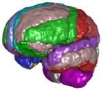
| - | [[Image: | + | [[Image:SOCR_Data_Dinov_July2009_NI.png|150px|thumbnail|right| Neuroimaging Data ]] |
| - | The [http:// | + | [[SOCR_Data_July2009_ID_NI#References |This is a large neuroimaging study]] using automated volumetric data processing to obtain different shape and volume measures of local anatomy. The subject population is derived from the [http://www.loni.ucla.edu/ADNI Alzheimer's Disease Neuroimaging Initiative (ADNI) database] and includes 27 Alzheimer's disease (AD) subjects, 35 normal controls (NC), and 42 mild cognitive impairment subjects (MCI). The goals of the study were to identify associations and relationships between neuroimaging biomarkers and various subject demographics and traits. |
==Data Description== | ==Data Description== | ||
| - | * '' | + | * Subject_ID: index of the subject in this dataset (1, 2, 3, ..., 104) |
| - | * ' | + | * Group: Indicator of the subject group: Alzheimer's disease (AD), normal control (NC), or mild cognitive impairment (MCI) |
| - | * ''' | + | * MMSE: [http://en.wikipedia.org/wiki/Mini-mental_state_examination Mini-Mental State Exam score], a cognitive assessment measure |
| - | * '' | + | * CDR: [http://en.wikipedia.org/wiki/Clinical_Dementia_Rating Clinical Dementia Rating scale] |
| - | * ''' | + | * Sex: Subject's gender label |
| - | * ''' | + | * Age: Subject's age |
| + | * TBV: Total Brain Volume measure in ''mm<sum>3</sup>'' | ||
| + | * GMV: Gray Matter Volume in ''mm<sum>3</sup>'' | ||
| + | * WMV: White Matter Volume in ''mm<sum>3</sup>'' | ||
| + | * CSFV: Cerebrospinal Fluid Volume in ''mm<sum>3</sup>'' | ||
| + | * ROI: [[SOCR_Data_July2009_ID_NI#ROIs |Label/Index of a Region of Interest (ROI)]] | ||
| + | * Measure: Labels of ROI measures: | ||
| + | ** SA: Surface Area of the ROI | ||
| + | ** SI: Shape Index of the ROI | ||
| + | ** CV: Curvedness of the ROI | ||
| + | ** FD: Fractal Dimension of the ROI | ||
| + | * Value: Numerical value of the measure (above). | ||
| + | |||
| + | |||
| + | ==Data== | ||
| + | [http://socr.ucla.edu/docs/resources/SOCR_Data/AD_NC_MCI_NI_Data2009.html Complete data set (5MB) is available here]. A small portion of the data that illustrates the complexity of this dataset is shown in the table below: | ||
| - | |||
{| class="wikitable" style="text-align:center;" border="2" | {| class="wikitable" style="text-align:center;" border="2" | ||
|- | |- | ||
| - | ! || | + | ! Subject_ID || Group || MMSE || CDR || Sex || Age || TBV || GMV || WMV || CSFV || ROI || Measure || Value |
| + | |||
| + | |} | ||
| + | |||
| + | == ROIs== | ||
| + | {| class="wikitable" style="text-align:center;" border="2" | ||
| + | |- | ||
| + | ! Index || ImageIntensity || ROI_Name | ||
| + | |- | ||
| + | | 0 || 0 || Background | ||
| + | |- | ||
| + | | 1 || 21 || L_superior_frontal_gyrus | ||
| + | |- | ||
| + | | 2 || 24 || R_middle_frontal_gyrus | ||
| + | |- | ||
| + | | 3 || 50 || R_precuneus | ||
| + | |- | ||
| + | | 4 || 181 || cerebellum | ||
| + | |- | ||
| + | | 5 || 47 || L_angular_gyrus | ||
| + | |- | ||
| + | | 6 || 122 || R_cingulate_gyrus | ||
| + | |- | ||
| + | | 7 || 83 || L_middle_temporal_gyrus | ||
| + | |- | ||
| + | | 8 || 90 || R_lingual_gyrus | ||
| + | |- | ||
| + | | 9 || 81 || L_superior_temporal_gyrus | ||
| + | |- | ||
| + | | 10 || 91 || L_fusiform_gyrus | ||
| + | |- | ||
| + | | 11 || 44 || R_superior_parietal_gyrus | ||
| + | |- | ||
| + | | 12 || 66 || R_inferior_occipital_gyrus | ||
| + | |- | ||
| + | | 13 || 87 || L_parahippocampal_gyrus | ||
| + | |- | ||
| + | | 14 || 162 || R_caudate | ||
| + | |- | ||
| + | | 15 || 85 || L_inferior_temporal_gyrus | ||
| + | |- | ||
| + | | 16 || 182 || brainstem | ||
| + | |- | ||
| + | | 17 || 43 || L_superior_parietal_gyrus | ||
| + | |- | ||
| + | | 18 || 28 || R_precentral_gyrus | ||
| + | |- | ||
| + | | 19 || 23 || L_middle_frontal_gyrus | ||
| + | |- | ||
| + | | 20 || 89 || L_lingual_gyrus | ||
| + | |- | ||
| + | | 21 || 41 || L_postcentral_gyrus | ||
| + | |- | ||
| + | | 22 || 86 || R_inferior_temporal_gyrus | ||
| + | |- | ||
| + | | 23 || 163 || L_putamen | ||
| + | |- | ||
| + | | 24 || 26 || R_inferior_frontal_gyrus | ||
| + | |- | ||
| + | | 25 || 102 || R_insular_cortex | ||
| + | |- | ||
| + | | 26 || 25 || L_inferior_frontal_gyrus | ||
| + | |- | ||
| + | | 27 || 46 || R_supramarginal_gyrus | ||
| + | |- | ||
| + | | 28 || 34 || R_gyrus_rectus | ||
| + | |- | ||
| + | | 29 || 65 || L_inferior_occipital_gyrus | ||
| + | |- | ||
| + | | 30 || 164 || R_putamen | ||
| + | |- | ||
| + | | 31 || 61 || L_superior_occipital_gyrus | ||
| + | |- | ||
| + | | 32 || 30 || R_middle_orbitofrontal_gyrus | ||
| + | |- | ||
| + | | 33 || 42 || R_postcentral_gyrus | ||
| + | |- | ||
| + | | 34 || 27 || L_precentral_gyrus | ||
| + | |- | ||
| + | | 35 || 32 || R_lateral_orbitofrontal_gyrus | ||
| + | |- | ||
| + | | 36 || 121 || L_cingulate_gyrus | ||
| + | |- | ||
| + | | 37 || 31 || L_lateral_orbitofrontal_gyrus | ||
| + | |- | ||
| + | | 38 || 92 || R_fusiform_gyrus | ||
| + | |- | ||
| + | | 39 || 45 || L_supramarginal_gyrus | ||
| + | |- | ||
| + | | 40 || 88 || R_parahippocampal_gyrus | ||
| + | |- | ||
| + | | 41 || 22 || R_superior_frontal_gyrus | ||
| + | |- | ||
| + | | 42 || 29 || L_middle_orbitofrontal_gyrus | ||
| + | |- | ||
| + | | 43 || 68 || R_cuneus | ||
| + | |- | ||
| + | | 44 || 62 || R_superior_occipital_gyrus | ||
| + | |- | ||
| + | | 45 || 33 || L_gyrus_rectus | ||
| + | |- | ||
| + | | 46 || 48 || R_angular_gyrus | ||
| + | |- | ||
| + | | 47 || 64 || R_middle_occipital_gyrus | ||
| + | |- | ||
| + | | 48 || 84 || R_middle_temporal_gyrus | ||
| + | |- | ||
| + | | 49 || 49 || L_precuneus | ||
| + | |- | ||
| + | | 50 || 67 || L_cuneus | ||
| + | |- | ||
| + | | 51 || 161 || L_caudate | ||
| + | |- | ||
| + | | 52 || 165 || L_hippocampus | ||
| + | |- | ||
| + | | 53 || 166 || R_hippocampus | ||
| + | |- | ||
| + | | 54 || 82 || R_superior_temporal_gyrus | ||
| + | |- | ||
| + | | 55 || 63 || L_middle_occipital_gyrus | ||
| + | |- | ||
| + | | 56 || 101 || L_insular_cortex | ||
|} | |} | ||
== References== | == References== | ||
* Dinov ID, Van Horn JD, Lozev KM, Magsipoc R, Petrosyan P, Liu Z, MacKenzie-Graha A, Eggert P, Parker DS and Toga AW (2009) Efficient, Distributed and Interactive Neuroimaging Data Analysis using the LONI Pipeline. [http://www.frontiersin.org/neuroinformatics/ Front. Neuroinform.] (2009) 3:22. [http://www.frontiersin.org/neuroinformatics/paper/10.3389/neuro.11/022.2009/ doi:10.3389/neuro.11.022.2009], published online: 20 July 2009. | * Dinov ID, Van Horn JD, Lozev KM, Magsipoc R, Petrosyan P, Liu Z, MacKenzie-Graha A, Eggert P, Parker DS and Toga AW (2009) Efficient, Distributed and Interactive Neuroimaging Data Analysis using the LONI Pipeline. [http://www.frontiersin.org/neuroinformatics/ Front. Neuroinform.] (2009) 3:22. [http://www.frontiersin.org/neuroinformatics/paper/10.3389/neuro.11/022.2009/ doi:10.3389/neuro.11.022.2009], published online: 20 July 2009. | ||
| + | |||
| + | * David W. Shattuck, Mubeena Mirza, Vitria Adisetiyo, Cornelius Hojatkashani, Georges Salamon, Katherine L. Narr, Russell A. Poldrack, Robert M. Bilder, Arthur W. Toga, Construction of a 3D probabilistic atlas of human cortical structures, NeuroImage, Volume 39, Issue 3, 1 February 2008, Pages 1064-1080, ISSN 1053-8119, DOI: [http://dx.doi.org/10.1016/j.neuroimage.2007.09.031 10.1016/j.neuroimage.2007.09.031]. | ||
| + | |||
<hr> | <hr> | ||
{{translate|pageName=http://wiki.stat.ucla.edu/socr/index.php?title=SOCR_Data_July2009_ID_NI}} | {{translate|pageName=http://wiki.stat.ucla.edu/socr/index.php?title=SOCR_Data_July2009_ID_NI}} | ||
Revision as of 05:55, 24 July 2009
Contents |
SOCR Datasets - Neuroimaging study of 27 Alzheimer's disease (AD) subjects, 35 normal controls (NC), and 42 mild cognitive impairment subjects (MCI)
Data Overview
This is a large neuroimaging study using automated volumetric data processing to obtain different shape and volume measures of local anatomy. The subject population is derived from the Alzheimer's Disease Neuroimaging Initiative (ADNI) database and includes 27 Alzheimer's disease (AD) subjects, 35 normal controls (NC), and 42 mild cognitive impairment subjects (MCI). The goals of the study were to identify associations and relationships between neuroimaging biomarkers and various subject demographics and traits.
Data Description
- Subject_ID: index of the subject in this dataset (1, 2, 3, ..., 104)
- Group: Indicator of the subject group: Alzheimer's disease (AD), normal control (NC), or mild cognitive impairment (MCI)
- MMSE: Mini-Mental State Exam score, a cognitive assessment measure
- CDR: Clinical Dementia Rating scale
- Sex: Subject's gender label
- Age: Subject's age
- TBV: Total Brain Volume measure in mm<sum>3</sup>
- GMV: Gray Matter Volume in mm<sum>3</sup>
- WMV: White Matter Volume in mm<sum>3</sup>
- CSFV: Cerebrospinal Fluid Volume in mm<sum>3</sup>
- ROI: Label/Index of a Region of Interest (ROI)
- Measure: Labels of ROI measures:
- SA: Surface Area of the ROI
- SI: Shape Index of the ROI
- CV: Curvedness of the ROI
- FD: Fractal Dimension of the ROI
- Value: Numerical value of the measure (above).
Data
Complete data set (5MB) is available here. A small portion of the data that illustrates the complexity of this dataset is shown in the table below:
| Subject_ID | Group | MMSE | CDR | Sex | Age | TBV | GMV | WMV | CSFV | ROI | Measure | Value
|
|---|
ROIs
| Index | ImageIntensity | ROI_Name |
|---|---|---|
| 0 | 0 | Background |
| 1 | 21 | L_superior_frontal_gyrus |
| 2 | 24 | R_middle_frontal_gyrus |
| 3 | 50 | R_precuneus |
| 4 | 181 | cerebellum |
| 5 | 47 | L_angular_gyrus |
| 6 | 122 | R_cingulate_gyrus |
| 7 | 83 | L_middle_temporal_gyrus |
| 8 | 90 | R_lingual_gyrus |
| 9 | 81 | L_superior_temporal_gyrus |
| 10 | 91 | L_fusiform_gyrus |
| 11 | 44 | R_superior_parietal_gyrus |
| 12 | 66 | R_inferior_occipital_gyrus |
| 13 | 87 | L_parahippocampal_gyrus |
| 14 | 162 | R_caudate |
| 15 | 85 | L_inferior_temporal_gyrus |
| 16 | 182 | brainstem |
| 17 | 43 | L_superior_parietal_gyrus |
| 18 | 28 | R_precentral_gyrus |
| 19 | 23 | L_middle_frontal_gyrus |
| 20 | 89 | L_lingual_gyrus |
| 21 | 41 | L_postcentral_gyrus |
| 22 | 86 | R_inferior_temporal_gyrus |
| 23 | 163 | L_putamen |
| 24 | 26 | R_inferior_frontal_gyrus |
| 25 | 102 | R_insular_cortex |
| 26 | 25 | L_inferior_frontal_gyrus |
| 27 | 46 | R_supramarginal_gyrus |
| 28 | 34 | R_gyrus_rectus |
| 29 | 65 | L_inferior_occipital_gyrus |
| 30 | 164 | R_putamen |
| 31 | 61 | L_superior_occipital_gyrus |
| 32 | 30 | R_middle_orbitofrontal_gyrus |
| 33 | 42 | R_postcentral_gyrus |
| 34 | 27 | L_precentral_gyrus |
| 35 | 32 | R_lateral_orbitofrontal_gyrus |
| 36 | 121 | L_cingulate_gyrus |
| 37 | 31 | L_lateral_orbitofrontal_gyrus |
| 38 | 92 | R_fusiform_gyrus |
| 39 | 45 | L_supramarginal_gyrus |
| 40 | 88 | R_parahippocampal_gyrus |
| 41 | 22 | R_superior_frontal_gyrus |
| 42 | 29 | L_middle_orbitofrontal_gyrus |
| 43 | 68 | R_cuneus |
| 44 | 62 | R_superior_occipital_gyrus |
| 45 | 33 | L_gyrus_rectus |
| 46 | 48 | R_angular_gyrus |
| 47 | 64 | R_middle_occipital_gyrus |
| 48 | 84 | R_middle_temporal_gyrus |
| 49 | 49 | L_precuneus |
| 50 | 67 | L_cuneus |
| 51 | 161 | L_caudate |
| 52 | 165 | L_hippocampus |
| 53 | 166 | R_hippocampus |
| 54 | 82 | R_superior_temporal_gyrus |
| 55 | 63 | L_middle_occipital_gyrus |
| 56 | 101 | L_insular_cortex |
References
- Dinov ID, Van Horn JD, Lozev KM, Magsipoc R, Petrosyan P, Liu Z, MacKenzie-Graha A, Eggert P, Parker DS and Toga AW (2009) Efficient, Distributed and Interactive Neuroimaging Data Analysis using the LONI Pipeline. Front. Neuroinform. (2009) 3:22. doi:10.3389/neuro.11.022.2009, published online: 20 July 2009.
- David W. Shattuck, Mubeena Mirza, Vitria Adisetiyo, Cornelius Hojatkashani, Georges Salamon, Katherine L. Narr, Russell A. Poldrack, Robert M. Bilder, Arthur W. Toga, Construction of a 3D probabilistic atlas of human cortical structures, NeuroImage, Volume 39, Issue 3, 1 February 2008, Pages 1064-1080, ISSN 1053-8119, DOI: 10.1016/j.neuroimage.2007.09.031.
Translate this page:

